Histology and Morphogenesis Laboratory: відмінності між версіями
Створена сторінка: Work with the snow-type microtome MS-2 | mini Working area of the histology and morphogenesis laboratory | mini Zone for microscopy and morphological description | mini Primo Star 3 laboratory microscope | mini [[Файл:Мазок крові, моноцит.jpg| Human blood (smear), monocyte. Romanovsky-Giemsa stain | mini]... |
Немає опису редагування |
||
| Рядок 1: | Рядок 1: | ||
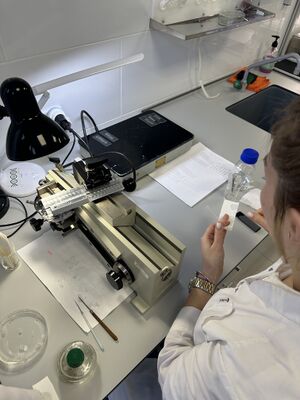
[[File:IMG 8322.jpg| Work with the snow-type microtome MS-2 | | [[File:IMG 8322.jpg| Work with the snow-type microtome MS-2|міні]] | ||
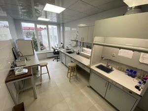
[[File:Photo 5359684286065791602 y.jpg| Working area of the histology and morphogenesis laboratory | | [[File:Photo 5359684286065791602 y.jpg| Working area of the histology and morphogenesis laboratory|міні]] | ||
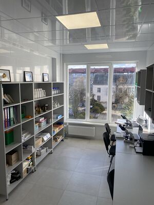
[[File:Photo 5359684286065791603 y.jpg| Zone for microscopy and morphological description | | [[File:Photo 5359684286065791603 y.jpg| Zone for microscopy and morphological description|міні]] | ||
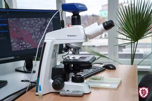
[[File:PrimoStar3.jpeg| Primo Star 3 laboratory microscope | | [[File:PrimoStar3.jpeg| Primo Star 3 laboratory microscope|міні]] | ||
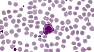
[[Файл:Мазок крові, моноцит.jpg| Human blood (smear), monocyte. Romanovsky-Giemsa stain | | [[Файл:Мазок крові, моноцит.jpg| Human blood (smear), monocyte. Romanovsky-Giemsa stain|міні]] | ||
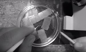
[[File:Petri.jpg| Video of histological preparation production https://www.youtube.com/watch?v=cjYZ0GTD5BA&t=2s | | [[File:Petri.jpg| Video of histological preparation production https://www.youtube.com/watch?v=cjYZ0GTD5BA&t=2s |міні]] | ||
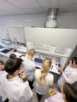
[[Файл:Фарбування з верху.jpg| Laboratory work "Making histological preparations" (specifically staining according to standard histological methods) with students of specialty 222 Medicine | | [[Файл:Фарбування з верху.jpg| Laboratory work "Making histological preparations" (specifically staining according to standard histological methods) with students of specialty 222 Medicine|міні]] | ||
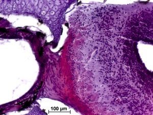
[[File:SNAP-121014-0011.jpg| Olfactory bulbs of the lake sturgeon. Hematoxylin and eosin | | [[File:SNAP-121014-0011.jpg| Olfactory bulbs of the lake sturgeon. Hematoxylin and eosin|міні]] | ||
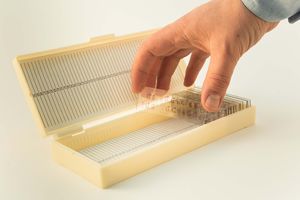
[[File:preparat.jpg| Archive of laboratory micropreparations | | [[File:preparat.jpg| Archive of laboratory micropreparations|міні]] | ||
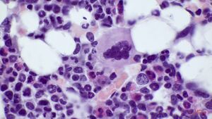
[[Файл: ЧКМ.Мегакаріоцит.jpg| Red bone marrow, megakaryocyte. Romanovsky-Giemsa stain | | [[Файл: ЧКМ.Мегакаріоцит.jpg| Red bone marrow, megakaryocyte. Romanovsky-Giemsa stain |міні]] | ||
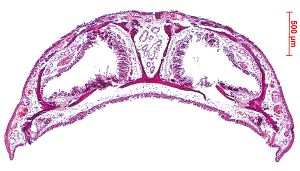
[[File:Ancistrus_dolichopterus.jpg| Cross-section of the head of the pseudolarva of Ancistrus dolichopterus through the olfactory rosette. Hematoxylin and eosin | | [[File:Ancistrus_dolichopterus.jpg| Cross-section of the head of the pseudolarva of Ancistrus dolichopterus through the olfactory rosette. Hematoxylin and eosin|міні]] | ||
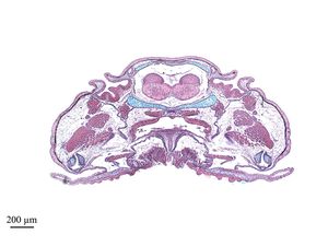
[[File:triturus.jpg| Cross-section of the head of the common newt. Hematoxylin and eosin | | [[File:triturus.jpg| Cross-section of the head of the common newt. Hematoxylin and eosin|міні]] | ||
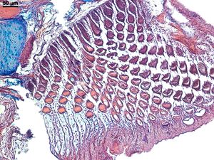
[[Файл:Поперечний переріз зубців ротової присоски псевдоличинки анциструса звичайного Ancistrus dolichopterus.jpg| Cross-section of the oral sucker teeth of the pseudolarva of the common Ancistrus dolichopterus (third place at the Scientific Photography Competition 2020) | | [[Файл:Поперечний переріз зубців ротової присоски псевдоличинки анциструса звичайного Ancistrus dolichopterus.jpg| Cross-section of the oral sucker teeth of the pseudolarva of the common Ancistrus dolichopterus (third place at the Scientific Photography Competition 2020) |міні]] | ||
The laboratory was established in 1993. | The laboratory was established in 1993. | ||
Версія за 09:11, 28 березня 2025
The laboratory was established in 1993. It operates at the Department of Histology and Medical Biology of the Medical Faculty.
Laboratory Documentation
Regulations on the Educational-Scientific Laboratory of Histology and Morphogenesis.
Program for introductory training.
Instruction No. 1 on BZD and labor protection issues.
Instruction No. 2 on providing first aid.
Instruction No. 13 on labor protection when working with computers.
Staff of the Laboratory
The laboratory employs:
Stepanyuk Yaroslav Vasilyovych, PhD in biology, associate professor, head of the laboratory;
Kostelova Olga Vasylivna, PhD in biology, senior lecturer;
Mironets Marina Yuriyivna, PhD student;
Pokotylo Olga Olegivna, PhD student.
Educational Components
Laboratory and practical sessions for students of the medical faculty, Faculty of Biology and Forestry and Chemistry, Ecology and Pharmacy are conducted at the laboratory:
Histology, Cytology, and Embryology (Assoc. Prof. Stepanyuk Y. V.)
Special Histology (Assoc. Prof. Stepanyuk Y. V.)
Cytology and Embryology with fundamentals of Teratology (Sr. Lect. Kostelova O. V., Assist. Mironets M. Yu.)
Current Issues in Developmental Biology (Sr. Lect. Kostelova O. V.)
Medical Biology (Assoc. Prof. Zinchenko M. O., Assoc. Prof. Stepanyuk Y. V.)
Methodologies
The laboratory uses the following methodologies:
Histological research methods;
Histochemical research methods;
Morphometric research methods (Zen ZEISS program);
3D reconstruction method based on serial micropreparations (Amira program);
Embryological research methods (experimental incubation of embryos of fish, amphibians, and reptiles).
Laboratory Equipment
Light Microscopes
ZEISS Primo Star 3 microscope with a color digital camera Axiocam 208 color;
MICROmed XS-5520 with a MIСmed 5 Mp video camera;
MicroBlue laboratory microscope;
Biolam R-1 microscope;
MB-10 stereomicroscope.
Laboratory Equipment
Snow-type microtome MS-2;
MOY-16 ocular micrometer;
Object micrometer;
Drying table for micropreparations;
Professional set for manual histological staining DiaPath;
Histological staining battery for micropreparations (Helenadechel cup set);
Drying oven, thermostats, scales, laboratory glassware, chemicals, dyes;
3D reconstruction equipment based on serial histological sections (computer, Wacom graphic tablet, software);
Specialized software for morphometric measurements;
Aquariums with equipment.
The laboratory has necessary collections of Scientific Collection of Histological Preparations and Educational Collection of Histological Preparations histological and cytological preparations.
Scientific Research Activities
Study of the morphology of the nervous system of vertebrates.
Morphogenesis and comparative morphology of the olfactory organ in vertebrates.
Features of the cytoarchitecture of the human olfactory bulbs.
Comparative morphology of the structures of the olfactory analyzer in micro- and macrosmatic species.
Scientific Clubs and Problem Groups
A Student Scientific Histological Club functions at the laboratory (leader - Assoc. Prof. Stepanyuk Yaroslav Vasilyovych).
Realized Scientific Projects
Morphogenesis of the olfactory organ in vertebrates 0120U101676;
Presidential Grant of Ukraine for supporting research by young scientists "Study of the morphogenesis of the vomeronasal organ and olfactory epithelium in amphibians (GP/F26/0193); [1];
Gans Collections and Charitable Fund for attending the "International Congress of Vertebrate Morphology" (Washington) [2];
Evolutionary-morphological aspects of ontogenetic variability and the diversity of life forms in vertebrates [http://nddkr.ukrintei.ua/view/rk/1c32110f1396a5bf23f1b114ce6e65d6 0106
Competitions and Awards
Photographs of histological micropreparations made in the laboratory have been awarded diplomas in the Science Photo Competition in Ukraine from Wikimedia Commons
- Diploma for 2nd place (2016)
- Diploma for 2nd place (2017)
- Diploma for 1st place (2019)
- Diploma for 3rd place (2020)
- Diploma for 2nd place (2021)
- Diploma for 3rd place (2022)
Collaboration with Other Institutions
In order to carry out joint scientific research and quality practical training for students, the laboratory has established cooperation with:
- Medical laboratory of LLC "GEMO MEDIKA Lutsk" (Cooperation agreement);
- KP "Volyn Regional Pathological and Anatomical Bureau" (Cooperation agreement);
- Rivne Research Expert-Criminalistic Center of the Ministry of Internal Affairs of Ukraine (Cooperation agreement);
- LLC "EXPERT PATHOMORPHOLOGICAL LABORATORY" (Cooperation agreement);
- Institute of Animal Biology of the National Academy of Agrarian Sciences of Ukraine (Cooperation agreement);
- Lviv National University of Veterinary Medicine and Biotechnologies named after S. Z. Hzhytskyi (Cooperation agreement).
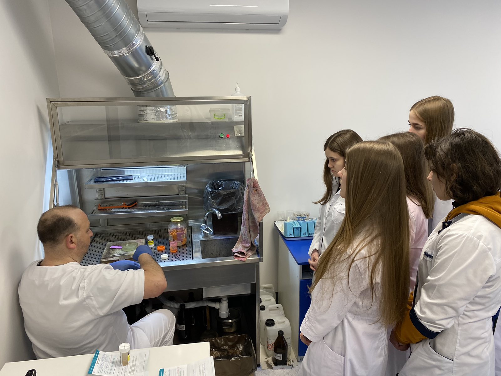

Scientific Events and Career Guidance Activities
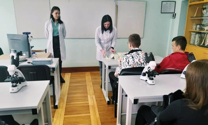
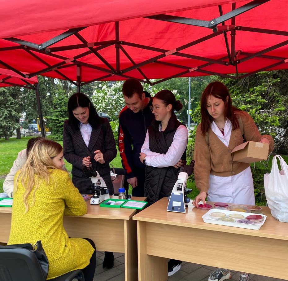
- On March 21, 2023, the training workshop "Day with Scientists" was held, where attendees of the regional Nature School, "Young Doctors" branch of the Volyn Ecological and Naturalistic Center had the opportunity to communicate with scientists and learn about the research activities of the Educational and Scientific Laboratory of Histology and Morphogenesis.
- On May 18, 2023, laboratory staff actively participated in the event during the Medical Faculty Week "HEALTH CAMP" - university camp: "Health and Mental Well-being: How to Take Care of Yourself During the War".
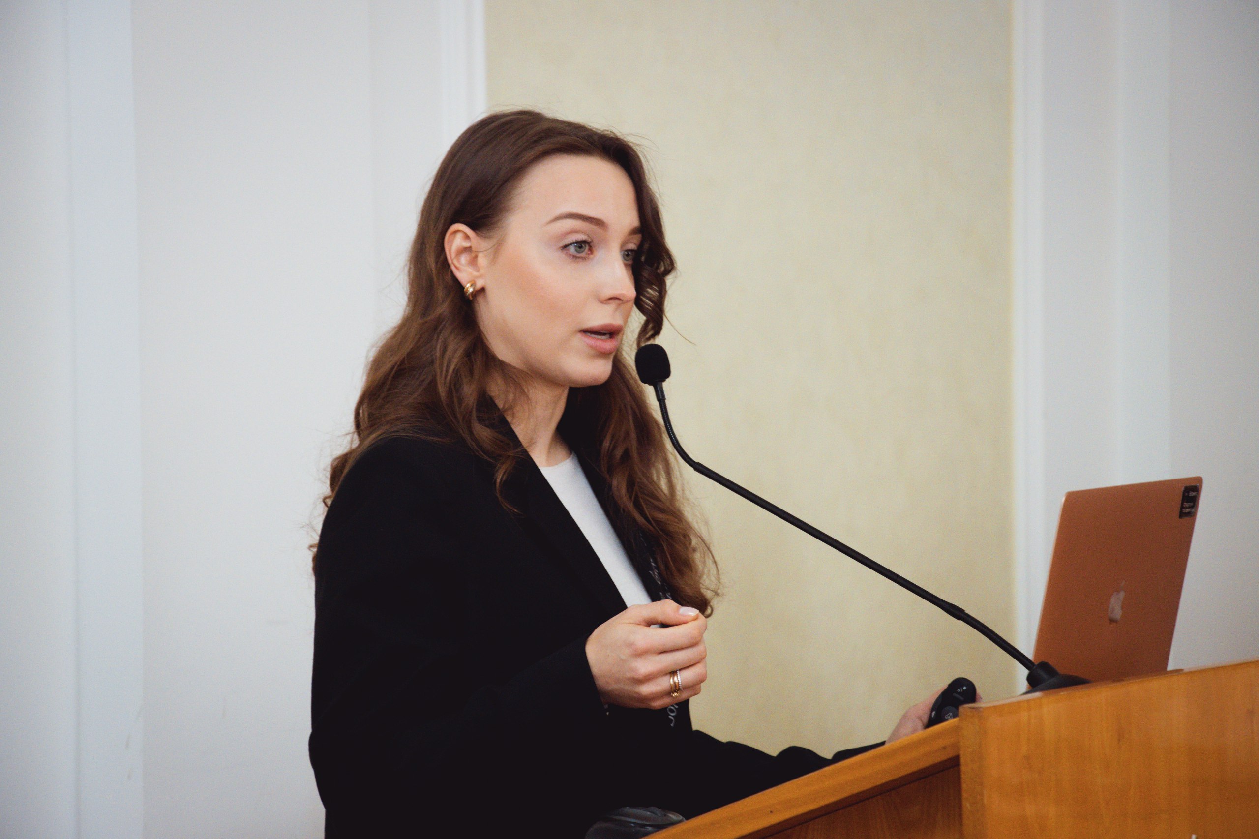
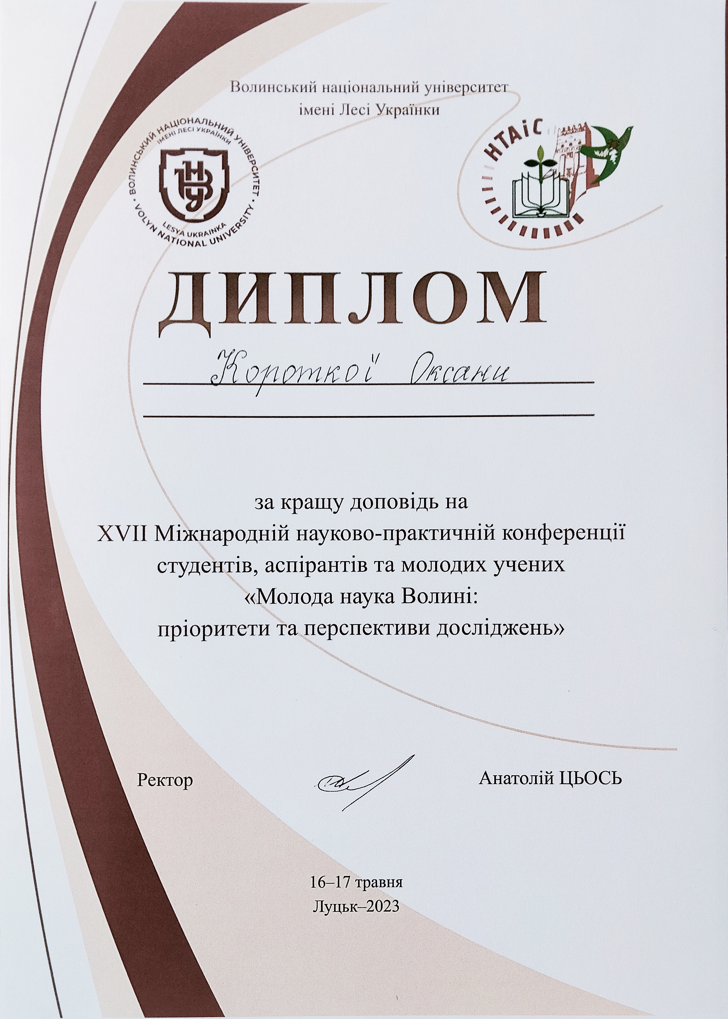
- Student Kоротка Оксана, a member of the student scientific histological club, gave a presentation "Modern Methods of Diagnosis and Treatment of Skin Diseases. A Morphologist's Perspective" at the plenary session of the XVII International Scientific and Practical Conference of Students and Postgraduates "Young Science of Volyn: Priorities and Prospects of Research".
Scientific Publications
- Pokotylo O., Mironets M., Stepanyuk Y. The history of the study of olfactory bulbs. "Young Science of Volyn: Priorities and Prospects of Research": materials of the XVII International Scientific and Practical Conference of Postgraduates and Students (May 16–17, 2023). Lutsk. Lesya Ukrainka Volyn National University, 2023. Pp. 675-678[3]
- Pokotylo O. O., Stepanyuk Y. V. Features of the cytoarchitecture of the human olfactory bulbs. Current Problems of the Development of Natural and Humanitarian Sciences: materials of the VI International Scientific and Practical Conference of Young Scientists, Students, and Postgraduates (Lutsk, November 11, 2022). Lutsk, 2022. P. 459–460. [4]
- Stepanyuk Y. V., Ulyanov V. O. The use of the 3D reconstruction method using the example of the development of the olfactory analyzer. Theory and Practice of Modern Morphology: materials of the Sixth All-Ukrainian Scientific and Practical Conference with international participation (Dnipro, November 9–11, 2022). Dnipro, 2022. P. 141. [5]
- Stepanyuk Y. V., Ulyanov V. O., Solovey L. M. Experience in the use of ZEN (ZEISS) software in teaching morphological educational components. Modern Concepts of Teaching Natural Sciences in Medical Educational Institutions: materials of the XV International Scientific and Methodological Online Conference (Kharkiv, November 15–16, 2022). Kharkiv: KhNMU, 2022. P. 40–41. [6]
Contact Information
- Lutsk, Bankova St., 9, S-322.
- email: Stepanyuk.Yaroslav@vnu.edu.ua
- email: Myronets.Maryna@vnu.edu.ua
- Department of Histology and Medical Biology profile on [7]
- Working hours of the Educational and Scientific Laboratory of Histology and Morphogenesis:
| Monday | 8.15 - 12.00 | 12.48 -17.15 |
| Tuesday | 8.15 - 12.00 | 12.48 -17.15 |
| Wednesday | 8.15 - 12.00 | 12.48 -17.15 |
| Thursday | 8.15 - 12.00 | 12.48 -17.15 |
| Friday | 8.15 - 12.00 | 12.48 -16.15 |
| Saturday | Day off | |
| Sunday | Day off | |
